Cavities, the stealthy culprits in dental health, often leave us wondering: what do cavities look like on an x ray? This guide not only answers that question but also delves into various facets, from the inception of cavities to reading dental X-rays for cavities. Let’s embark on a journey into the world of your teeth and the secrets an X-ray can unveil.
- Unveiling the Early Stages: What Does a Cavity Look Like When It First Starts?
- The Big Picture: Big Cavity X-Ray
- Deciphering the Code: How to Read Dental X-Rays for Cavities
- The Filling Chronicles: What Does a Filling Look Like on an X-Ray?
- Journey Inside: What Does a Cavity Look Like Inside?
- Side Stories: What Does a Cavity Look Like on the Side of a Tooth?
- Contrast Chronicles: Healthy vs. Unhealthy Teeth X-Ray
- Navigating Discomfort: Do Cavities Hurt?
- Empowering Your Oral Health Journey
Unveiling the Early Stages: What Does a Cavity Look Like When It First Starts?
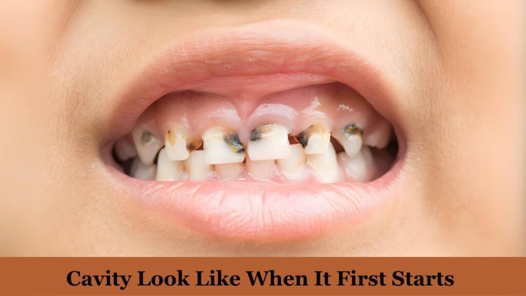
Before we dive into X-ray revelations, let’s explore the sneak peek into the early stages of cavity development. What does a cavity look like when it first starts? In its infancy, a cavity manifests as a subtle darkness or radiolucent area within the enamel on an X-ray. This faint shadow signals the beginning of a potential dental foe.
Understanding the genesis of cavities is crucial. Imagine it as the opening scene of a suspenseful movie – a small, inconspicuous anomaly that, if unnoticed, could evolve into a major plot twist. The X-ray, your cinematic tool, captures this initial stage, providing a narrative that guides your dental health story.
The Big Picture: Big Cavity X-Ray
As cavities progress, they may grow in size, leading to the dreaded big cavity scenario. An X-ray of a sizable cavity presents a prominent radiolucent area, revealing the extent of tooth decay. Understanding the visual cues in a big cavity X-ray can aid in comprehending the severity of the condition.
In this stage, the plot thickens. The X-ray transforms into a panoramic view, showcasing the impact of neglect or oversight. Picture a vast landscape with dark valleys, signifying the areas where decay has taken hold. The big cavity X-ray serves as a cautionary tale – a reminder that proactive measures are vital in preventing the escalation of dental challenges.
Deciphering the Code: How to Read Dental X-Rays for Cavities
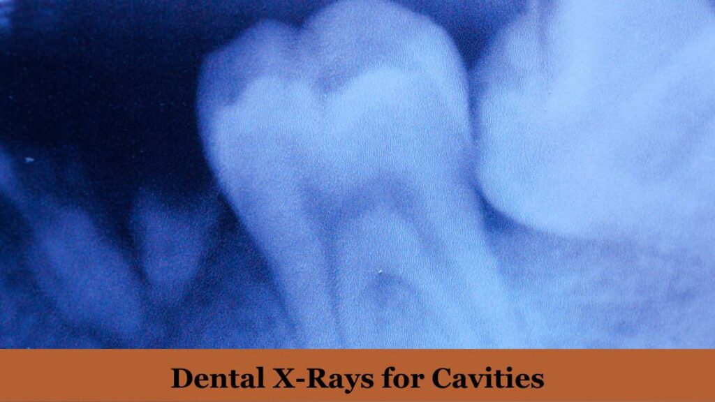
Reading dental X-rays for cavities requires a bit of decoding. This section guides you through the process, breaking down the radiopaque and radiolucent language. Learn the art of identifying cavities in different areas – from the chewing surface to underneath fillings. Mastering how to read dental X-rays for cavities empowers you to actively participate in your oral health journey.
Think of this decoding process as unraveling a mystery. Each X-ray is a unique puzzle, and deciphering the code is akin to solving clues. The radiopaque structures are the visible, well-preserved elements, while radiolucent areas mark the spots where the plot thickens – where cavities silently make their presence known. This newfound skill transforms you into a dental detective, equipped to preemptively address potential oral health challenges.
The Filling Chronicles: What Does a Filling Look Like on an X-Ray?
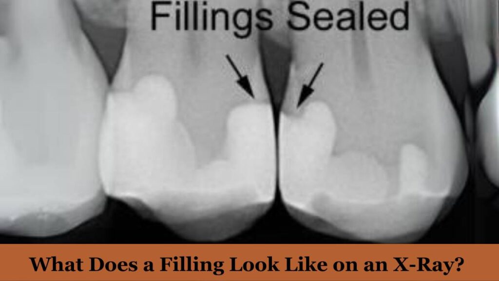
Ever wondered how a dental filling appears on an X-ray? This segment demystifies the imagery, illustrating what a filling looks like within the tooth structure. Uncover the intricacies of what does a filling look like on an X-ray and grasp the transformative role it plays in preserving your tooth’s integrity.
As the narrative progresses, the filling emerges as a crucial character in your dental story. The X-ray captures its presence – a solid, radiopaque entity strategically placed to restore balance. It’s the turning point where intervention meets resolution. The filling, a guardian of dental harmony, becomes a beacon of hope in the radiographic landscape.
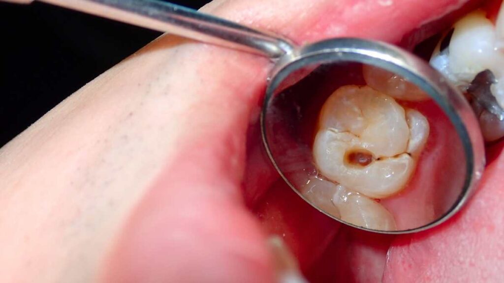
Journey Inside: What Does a Cavity Look Like Inside?
Venture into the depths of a tooth to explore what a cavity looks like inside. Beyond the surface, an X-ray unravels the internal landscape of a cavity, providing valuable insights into its size, shape, and potential impact on surrounding structures. Knowledge is power, and understanding the internal dynamics equips you to make informed decisions about your dental care.
This journey inside is akin to exploring hidden chambers in a mystical realm. The X-ray acts as your magical map, revealing the caverns where decay has taken residence. Each shadow, each radiolucent contour, tells a story of vulnerability and resilience. Armed with this knowledge, you become the architect of your dental fate, deciding the course of action to preserve the sanctity of your dental kingdom.
Side Stories: What Does a Cavity Look Like on the Side of a Tooth?
Cavities don’t always take center stage; sometimes, they linger on the sidelines. Explore what a cavity looks like on the side of a tooth through X-ray imagery. Uncover the nuances of side-oriented decay, offering a comprehensive view of dental health from all angles.
In this narrative, the side story adds depth and dimension. It’s the subplot that, if ignored, could impact the overall arc of your dental journey. The X-ray captures these peripheral tales, urging you to pay attention to every facet of your dental landscape. The side of a tooth becomes a canvas, and cavities, mere brushstrokes that need careful consideration.
Contrast Chronicles: Healthy vs. Unhealthy Teeth X-Ray

To truly grasp the impact of cavities, comparing healthy and unhealthy teeth on an X-ray becomes essential. This section juxtaposes the radiographic differences, providing a visual journey through the healthy vs. unhealthy teeth X-ray spectrum. Witness the stark contrasts and appreciate the significance of preventive dental measures.
Imagine this section as a gallery – one wall adorned with vibrant, radiopaque portrayals of robust dental health, and the other depicting the shadows and voids of untreated cavities. The healthy vs. unhealthy teeth X-ray showcase underscores the importance of proactive measures. It’s a call to action, encouraging you to embrace the radiance of preventive dental care.
Navigating Discomfort: Do Cavities Hurt?
Amidst the visual exploration, let’s address a common concern – do cavities hurt? Understanding the correlation between cavity progression and discomfort is crucial. Gain insights into when and why cavities might cause pain, enhancing your awareness of potential oral health challenges.
This segment introduces a human element to the narrative. It explores the emotions tied to dental health, addressing the fear or discomfort that cavities may evoke. The X-ray, a diagnostic ally, assists in navigating this discomfort. It’s a tool that not only reveals the physical manifestations but also guides the emotional journey towards resolution.
Empowering Your Oral Health Journey
Armed with insights into what cavities look like on x rays and equipped with knowledge about their early stages, progression, and impact, you are now a proactive participant in your oral health journey. Remember, regular check-ups and a proactive approach to dental care are your allies in maintaining a radiant and healthy smile.
Note: This article is not a substitute for professional dental advice. Consult your dentist for personalized guidance.

A true master in his field, Hayate takes the helm as the author of Supreme Hall’s ‘Health’ category. Through his informative and engaging blogs, he shares valuable insights on wellness, fitness, and holistic living, empowering readers to lead healthier and happier lives.


Looks good for me. Thank Youh!